
Anatomy Chart courtesy of FCIT
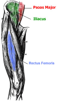
The Iliacus originates on the pelvic crest and attaches on the femur.
The Psoas Major, the longer of the two muscles, originates on the lumbar vertebrae and attaches to the femur.
The Rectus Femoris is also a hip flexor. It is one of the four Quadriceps muscles and the only one that crosses the hip joint. This crossing of the hip joint enables it to operate as a hip flexor as well as a knee extensor (straightening the knee).
Overdeveloped and tight hip flexors can contribute to lower back pain by causing the pelvis to tilt forward. To counteract this, you must stretch the hip flexors and strengthen the Abdominal muscles. This will reduce pelvic tilt and decrease lower back pain. Strengthening the lower back can also help improve the balance between the muscles of the hip region.